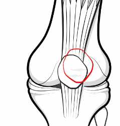
Patellar malalignment is present when the kneecap - for whatever reason - does not sit optimally in the underlying groove as the knee bends and straightens.
 Page updated July 2024 by Dr Sheila Strover (Clinical Editor)

Malalignment is an observation, not a diagnosis
Causes of patellar malalignment may be local or related to abnormal rotations at the hip or femur.
So malalignment is an observation or an inference, rather than a diagnosis, and the cause of any malignment needs to be sought in cases of recurrent anterior knee pain or when subluxation or dislocation of the patella occurs in the absence of any precipitating injury.
The patella may be too high
This condition is known as patella alta. When the knee bends, the patella is too high to easily engage in the groove of the underlying femur (trochlear groove), which is shallow in its upper end.
So the patella sometimes fails to engage and slips over the side, subluxing or dislocating. Patella alta is something one is born with, and if it is very problematic only surgery will correct the problem.
The patella may be tilted to one side
Patellar tilt is due to the fibrous tethers (retinacula) being too tight on one side and tugging the patella so that it tips over.
Usually it is the lateral retinaculum on the outer side of the knee that is too tight. This may be secondary to tightness in the iliotibial band or weakness of the VMO part of the quads muscle group. Appropriate exercise and patellar mobilisation stretches may help to improve the situation.
The patella may be an abnormal shape
An abnormally-shaped patella is called patellar dysplasia.
This is also something that one is born with. The rear of the patella may be abnormally flat, rather than V-shaped. Or the patella may be smaller than normal.
The groove under the patella may be abnormal
An abnormal groove in the femur is called trochlear dysplasia.
There are several degrees of dysplasia, but in the most extreme form the upper end of the groove actually bulges out rather than being concave. Trochlear dysplasia is something one is born with. Surgery to correct it is a super-specialist procedure.
The patellar tendon attachment may not be optimal
The patellar tendon attaches to the tibia bone via the lump called the tibial tubercle.
Sometimes this attachment is not optimal (one is born that way), and the patellar surgeon may correct the positioning by moving the tubercle upwards or sideways and fixing it in the new position with a screw until bony healing takes place, at which point the screw can be removed. Procedures include tibial tuberosity transfer and distalisation of the tibial tubercle.
The long bones may be abnormally rotated
Despite having a totally normal patella, patellar tendon and trochlear groove, the patella may appear mal-aligned if one or both of the femur or tibia are themselves abnormally rotated.
This is called femoral torsion or tibial torsion.
Forum discussions
- MPFL repair,lateral release, Patella alignment 284 days ago
Patient discussing a past patellar alignment procedure.
Relevant material -
- Patellar tilt
- Patella alta
- Trochlear dysplasia
- J-sign
- TT-TG distance
- Femoral anteversion
- Miserable malalignment
Peer-reviewed paper -
- Journal interpretation - 2016 - First time patellar dislocation in children – risk factors. Authors: Askenberger M et al. - and interpreted for you by Dr Sheila Strover (Clinical Editor)
 2013 -
  2012 -