Articular cartilage repair is the repair or improvement of defects of the joint cartilage, either by surgery or biological intervention, or both.
 Page updated May 2024 by Dr Sheila Strover (Clinical Editor)

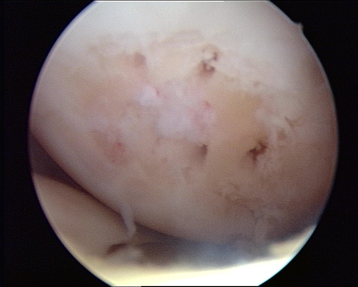
Articular cartilage injury on a weight-bearing part of the femoral condyle.
Articular cartilage injury in the knee
The knee is particularly vulnerable to articular cartilage damage, because of falls and direct blows during sporting activities.
Damage may range from a softening of the cartilage (chondromalacia), to a full thickness lesion (chondral defect), and a chunk of material may even be knocked off into the joint cavity (loose body).
-
Quote from peer-reviewed paper:
"Cartilage injuries can occur due to acute trauma or repetitive microtrauma....and this increases dramatically to 10–20 times of bodyweight during sports activities...Most early stage cartilage lesions do not cause a lot of symptoms and disability. However, in later stages, patients present with pain, swelling and mechanical symptoms like locking, catching and crepitus."
Citation: Solanki K, Shanmugasundaram S, Shetty N, Kim SJ. Articular cartilage repair & joint preservation: A review of the current status of biological approach. J Clin Orthop Trauma. 2021 Sep 21;22:101602. doi: 10.1016/j.jcot.2021.101602. PMID: 34631411; PMCID: PMC8488240.
How do you regenerate articular cartilage?
Articular cartilage is very different from most of the body's tissues, inasmuch as the cells are far apart from each other, suspended in a matrix without a proper blood supply.
Injury means that the cartilage does not heal well, and even where there is some healing the area tends to produce 'fibrocartilage' rather than true articular cartilage.
This has given rise to a number of special techniques involving bringing in blood components from the deeper sub-chondral layer, and also transferring healthy cells grown in a laboratory or tissue pieces transferred from other healthy areas where they are not so important.
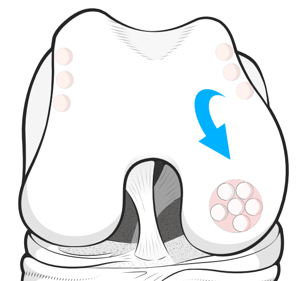
Microfracture to allow stem cells and nourishment
Microfracture for articular cartilage repair is a marrow stimulation technique, where holes are picked through the cartilage base plate, to allow blood and cells to track through into the defect.
The procedure may be performed in isolation, or in addition to another cartilage repair procedure.
Nanofracture is a variant of this original procedure, using smaller picks.
Cell multiplication in a laboratory
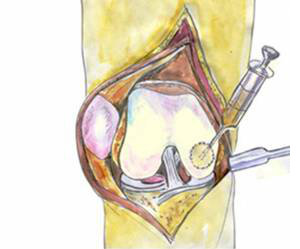
ACI techniques rely on cartilage cells being multiplied in the laboratory and being transferred to the damaged area, in this illustration being injected behind a membrane that has been sewn over the defect.
Transfer of healthy tissue from less important areas
Forum discussions
- Does BUPA cover MACI/ACI surgery
Patients discuss whether insurers will pay for cartilage repair procedures.
- exercise after OATS
Patients discuss how to protect the cartilage repair while rehabilitating.
- Microfracture
- Nanofracture
- Debridement
- Abrasion arthroplasty
- Marrow stimulation
- Osteochondral graft
- OATS
- Mosaicplasty
- Osteochondral allograft
Other relevant material -
- Autologous chondrocyte implantation (ACI)
- Matrix-induced Autologous Chondrocyte Implant (MACI)
- Orthobiologic
- Cartilage autograft implantation system (CAIS)
- Autologous matrix-induced chondrogenesis (AMIC)
- Nanofracture Autologous Matrix-Induced Chondrogenesis (NAMIC)
 2017 -
  2008 -
 eBook - Knee arthritis interventions when you are too young for a total knee replacement
Easy-to-read eBook discussing the various options for arthritis in the younger patient.