Knee anatomy includes all the various components of the knee, both inside and outside of the joint's waterproof capsule.
 Page updated June 2024 by Dr Sheila Strover (Clinical Editor)
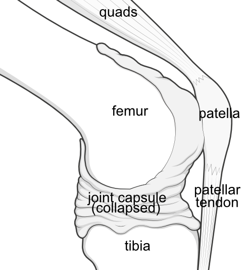
Side view - with the joint hidden within the deflated capsule.
The knee joint is not a simple hinge but a fundamentally unstable moving joint of three bones, enclosed within a stretchy watertight capsule.
The rest of the knee's anatomical complexity relates to all the other structures that stabilise these bones while still allowing them to move.
The bones of the knee
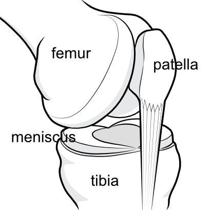
The three main bones of the knee are the femur (the thighbone), the tibia (the shinbone) and the patella (the kneecap).
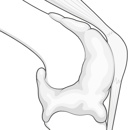
Side view - the bones within the canpsule.
The femur and tibia are the weight-bearing bones. The patella is different, and is actually embedded within the tendon of the large muscle that does much of the work of the knee.
Sometimes there is a small bone at the back of the joint, which mirrors the patella, but is tiny. It is called the fabella.
On the outer side of the knee, but outside of the capsule, is the fibula bone.
The capsule keeps the joint lubrication inside, where it is needed
The capsule is lined inside by a cell layer called the synovium which secretes the joint fluid that lubricates and nourishes the moving bony parts, and keeps their shiny, white joint surfaces healthy.
This joint surface is called articular cartilage, and it is key to understanding knee arthritis.
Side view - the capsule inflated with fluid.
Damage to the joint structures may lead to an excess of this synovial fluid, and the knee can blow up like a balloon (called an effusion). Sometimes an injury can lead to bleeding inside the capsule, again causing the knee to swell as the fluid cannot easily escape. This is called an haemarthrosis.
The big bones are linked and stabilised by the cruciate ligaments and menisci
The contact surfaces between femur and tibia are fundamentally unstable, because at its end the femur is rounded into two knuckles (condyles), while the top of tibia that they sit on is flat (tibial plateau).
So the incongruence between the shapes is resolved to some extent by crescent-shaped shock-absorbers wedged between them (menisci), while further stability is provided by ligaments holding the bones together (cruciate ligaments).
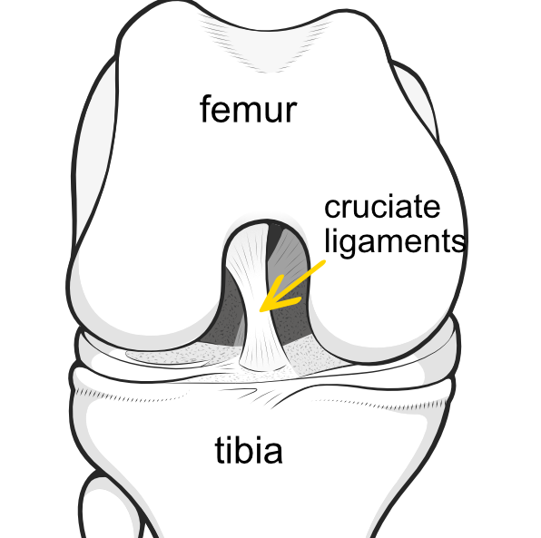
Front view - the capsule has been removed so that you can see the cruciate ligaments inside the joint.
The cruciate ligaments are contained within a notch at the bottom of the femur (femoral notch).

Oblique view - the cruciate ligaments have been removed to show how the patella slots into the groove of the femur (trochlear groove).
You can see the two crescentic menisci. The meniscus on the inner aspect of the knee is called the medial meniscus and the one on the outer aspect of the knee is called the lateral meniscus.
Further stability is provided on either side of the joint by the collateral ligaments (not illustrated).
The patella functions as a fulcrum
The patella exists within the tendon of the large muscle that makes up your 'lap' (quadriceps) and the tendon continues to attach on the tibia.
The part of the tendon above the patella is called the quadriceps tendon and the part below is called the patellar tendon, but it is really all one big tendon with the bone right inside it.
The patella is kept in its central position by the walls of the groove in the femur (trochlear groove). Besides being contained by the groove in the femur, the patella is further stabilised by a sheet of fibrous tissue on either side, including a strong band called the lateral retinaculum

Side view - to show how the patella is part of a pulley system.
Patella, muscle and the fibrous tissue together are called the extensor mechanism' because they work together to support the knee when it is straightened (extended). Thus contraction of the quads muscle will force the leg straight (into extension).
The muscles provide power
The quadriceps muscle group provides most of the power when straightening the knee (extension), while the hamstrings muscle group provides most of the power when bending the knee (flexion).
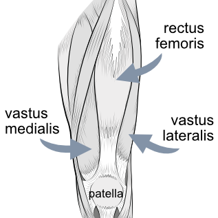
Front view - showing how the quadriceps muscle group relates to the patella.
When the thigh muscles at the front contract the calf muscles at the back relax, and vice versa.
The nerves relay information to the knee
Sensory nerves supply sensation to the knee, while motor nerves instruct the muscles to move.
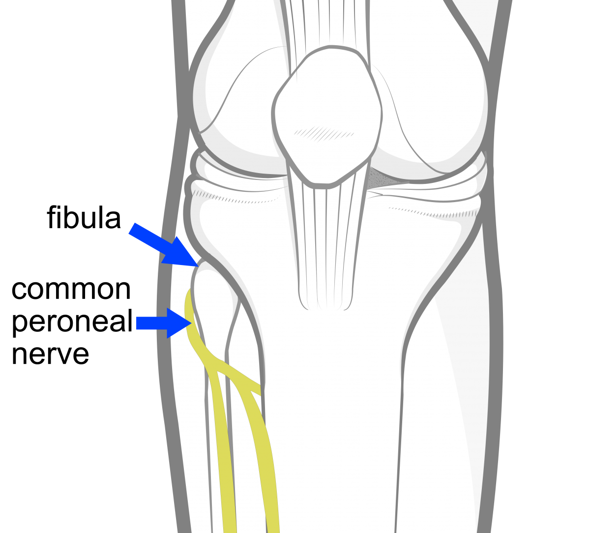
Front view - to show the important sciatic nerve.
The sciatic nerve splits just above the knee into the tibial nerve and the common peroneal nerve. The other important nerve is the saphenous nerve.