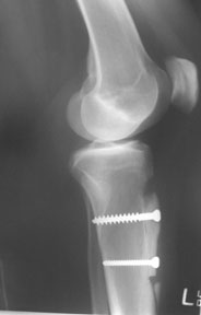
This is a course on limb realignment, but limb alignment also affects the patella.
 First published 2010, and reviewed August 2023 by Dr Sheila Strover (Clinical Editor)
Realignment osteotomy for knee pain - course
Part 1 - Introduction to the subject of knee osteotomy
Part 2 - Osteotomy for varus and valgus deformity
- Indications for varus and valgus osteotomy
- Living with painful varus and valgus deformity
- High tibial osteotomy and distal femoral osteotomy
- Case study of high tibial osteotomy aiding ligament instability
- Benefits of varus/valgus osteotomy
- Potential problems with varus/valgus osteotomy
- Recovery post knee osteotomy
Part 3 - Osteotomy for patellar instability
- Patellar instability and dislocation
- Dislocators with normal anatomy prior to dislocation
- Dislocators with abnormal anatomy prior to dislocation
The abnormal anatomy group is more intriguing for surgeons, and the first thing to do is to define the anatomical problems.
There are a number of specific anatomical abnormalities that we are on the lookout for -
Valgus alignment
Patients with abnormal anatomy are in valgus alignment, that is looking from the front, they really are quite knock-kneed. This means that the kneecap is already tending to slide off the lateral side of the knee.
Increased Q-angle
The patient often has what we call an ‘increased Q-angle’ (quadriceps angle). First you draw a straight line from the anterior superior iliac spine (the knobbly bit on the front of the pelvis) to the middle of the kneecap and down to the foot. And then you draw another line from the middle of the kneecap down the middle of your patellar tendon to your tibial tubercle, thereby creating an angle. In most people this angle is approximately 15 degrees. It is slightly greater in women than in men.
The Q-angle is one of those things that is very easy to talk about in clinic, it takes two seconds to do, but no-one actually draws lines and measures the angle with a goniometer.
Increased TTTD
These days we do have a very sophisticated way of measuring the Q angle and it is called the ‘tibial tubercle transfer distance’. Previously, we used CT scans to calculate this figure but that involved use of a lot of X-ray radiation. Now we can get the same information from an MRI scan (which does not involve any radiation). An MRI is made of digital slices and you can take the axial slices and use them to look at sections of the leg, like a tree that is being sliced from the top. The MRI can calculate very accurately how many millimetres it is from the middle of the trochlea to the middle of the tibial tubercle. Up to 18 mm is OK and anything over 20mm is really very abnorma. If the TTTD is over 20 mm and the patient is having serious problems with patella instability then surgery to reduce/normalise the TTTD is indicated.
J-tracking
Often when these patients sit with their leg dangling at 90 degrees of bend (flexion) and they straighten their leg up nice and slowly the kneecaps moves/tracks in an abnormal fashion. Instead of going up and down the leg (as you straighten and bend) it tracks towards the side and we call that J-tracking.
Hypermobility
When we examine these patients they are often hypermobile, that is all of their joints are a bit too mobile (thumb, metacarpophalangeal joints of the fingers, they hyperextend at the elbow, they hyperextend at the knee).
Age and gender
Usually these patients have had problems from a very early age. It is more common in females than in males, who may start to have trouble when they are 13 or 14 and occasionally even younger (the most common age is late teens or early twenties) They tend to complain of pain, and the pain comes from the instability.
Lateral tilt
When you look at the normal knee, the kneecap sits right over the front of the thigh. With someone with patellar instability usually their kneecap is actually tilting, pointing out to the outside.
Trochlear dysplasia
Once we have done our clinical assessment, we then do our radiological assessment and the critical X-ray view here is the lateral X-ray. From the lateral knee x-ray we can tell all sorts of things – we can tell the depth of the trochlear groove and we can tell its shape. In some individuals with a mis-shaped trochlea, developmental abnormalities can lead to either a flat or humped trochlea. The kneecap is therefore not sitting in a groove but sitting up on a hump. Both a flat and a humped trochlea fall under the umbrella of trochlear dysplasia. You can pick this up that on a lateral knee X-ray and this is really a critical investigation that can be carried out in in outpatients.
Patella alta
From the lateral X-ray again we then look critically at patellar height and we can see if the patella is low (patella baja or infera) or more commonly in these patients the patella is slightly high (patella alta).
So it is like building up on a points system, and when you get to a certain number of points you start running into trouble. So if you are a teenage girl, hypermobile, in valgus alignment with an increased Q-angle, it won't take very much for the kneecap to suddenly slide off the side and dislocate.
Subluxator or dislocator?
These are two very different groups. There are patients who actually dislocate and then there are those ones who subluxate (where the knee cap drops out and then drops back in again). A very key part of the assessment is asking the question “Have you ever been to casualty (ER) with your kneecap stuck to the side of your leg and it’s had to be put back in again?” If the answer is “yes”, then the patient may require the surgery. If the answer is “no, it always pops back in by itself”, then the patient is unlikely to require surgery. Usually these patients settle with appropriate physiotherapy.
Lateralised Tibial Tubercle
The other more common abnormal anatomy is when the trochlea is OK but the tibial tubercle is offset too far laterally. This can be addressed with the so-called Fulkerson osteotomy or modification of that particular procedure. Whatever you want to call it, and there are a number of different names, the idea is that you osteotomise the distal insertion of the patellar tendon (at the tibial tubercle) with a 4 cm block of bone and you transfer and fix it medially. The transfer distance is calculated on the MRI scan that you do pre-operatively. This operation has been given lots of different names and there have been lots of different variations on the original procedure.  The most commonly done operation now is a so-called modified Elmslie-Trillat or Fulkerson procedure. What that procedure does is it not only moves the tibial tubercle medially but the osteotomy is carried out in such a way that you move the bone slightly anteriorly so that not only are you pushing it medially but you are pushing it anteriorly. If you imagine if you cut the tibia from the lateral side if you cut it completely flat and you push the bone, the bone will just move in a flat plane. If you drop your hand and you cut it up a hill, you slide that piece of bone uphill. Conversely if you lift your hand up and you cut downhill, you can slide that piece of bone downhill. What we know is by dropping our hands and pushing it upwards we take pressure off the patellofemoral joint by moving everything anteriorly. If you do it flat it is ok, if you actually go downhill it is bad news because you are actually bringing too much pressure into the patellofemoral joint so what we tend to do is drop our hand from the lateral side, cut the bone in such a way that we slide the piece of bone uphill and then we fix it with a couple of screws. The problem with that procedure is that if you ask most surgeons “How far do you push that piece of bone?” you tend to get the same response and that is “A centimetre”.
Now obviously everyone is going to be slightly different. When I was working in Brisbane we researched a very novel idea that one of the surgeons, Peter Myers had and which he worked on with Andy Williams. Their idea was to stimulate the femoral nerve during the operation, which would make quadriceps muscle contract and pull on the patella, giving you some sort of feel for when the patella is in the middle of the trochlear groove.
Initially when you stimulate the femoral nerve the patella will jump off to the side laterally because it is tilted and it is sitting laterally. When you do your osteotomy and you get the patella to sit in the middle and then you stimulate the femoral nerve, the patella will not go too far laterally or go too far medially – it will tend to sit in the middle. And that was a very nice idea, but this is something that is quite difficult to achieve during the operation. But that is now how I do it – we do intraoperative femoral nerve stimulation and that gives us a slightly more scientific feel about how far we should be moving the tibial tubercle. It might be slightly imprecise but it works extremely well. This tends to be a bilateral problem and the patient will often ask: “When can you do my other side?”, so it is a very successful operation.
