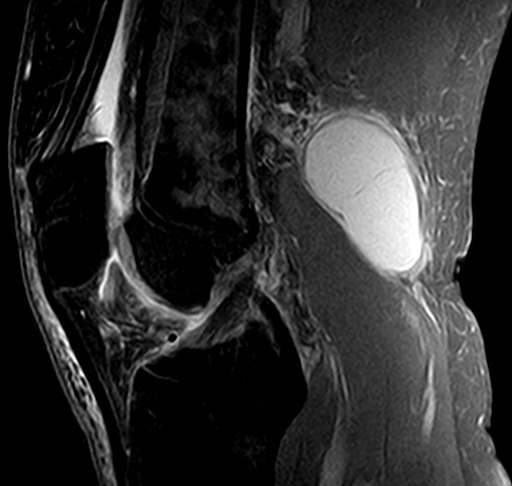
A Baker's cyst is a tense fluid-filled swelling at the back of the knee which is secondary to increased pressure of joint fluid inside the knee.
 Page updated June 2024 by Dr Sheila Strover (Clinical Editor)

A Baker's cyst can appear suddenly and be experienced as a painful lump on the inner aspect of the crease at the back of the knee on the inner aspect.

The cyst may become tense and rupture, with the fluid tracking into the back of the calf, mimicking a deep vein thrombosis.
What causes a Baker's cyst?

Increased fluid pressure inside the knee from some damage or irritation pushes against a weak area in the bursa (pocket) between the gastrocnemius and semimembranosus tendons of the muscles at the back of the knee.
The wall of the bursa can break down, allowing a valve-like flap that allows joint fluid to enter but not escape.
-
Quote from peer-reviewed paper:
"...commonly found in association with intra-articular knee disorders, such as osteoarthritis and meniscus tears....there may be chronic nonspecific inflammation present....loose bodies may also be found within the cyst. Baker’s cysts can be a source of posterior knee pain that persists despite surgical treatment of the intra-articular lesion...."
Citation: Frush TJ, Noyes FR. Baker's Cyst: Diagnostic and Surgical Considerations. Sports Health. 2015 Jul;7(4):359-65. doi: 10.1177/1941738113520130. PMID: 26137182; PMCID: PMC4481672.
How is a Baker's Cyst treated?
The surgeon usually initially focuses attention on the underlying cause of the cyst, checking for arthritis or a torn meniscus or any other problem inside the joint.
Resolution of the underlying cause may lead to decreased fluid pressure inside the knee, and the cyst may shrink. If this fails, some surgeons will aspirate the fluid with a syringe, although this may give only temporary relief. Arthroscopic surgery to remove the flap at the entrance to the cyst is becoming the recommended practice.
What happens if a Baker's cyst pops?
Rupture of a Baker's cyst may allow the fluid to track into the calf, where it can cause pain and swelling, and be confused with a deep vein thrombosis.
Fluid in the Baker's cyst shows up in white in this side-view MRI scan towards the right of the image. You can see its relationship to the gastrocnemius muscle which is just below it.
Forum discussions
- Knee brace for baker's cysts?
A discussion between patients about the management of a Baker's cyst.
- Pictures of my problem....Take a look and give me some ideas!
Patient photographs of the back of the knee.
Also relevant -
 2018 - Surgery for Baker´s Cyst - by Dr Lars Blønd (Knee Surgeon)
 2016 - Baker's Cyst - Symptoms and Causes - by Dr Sheila Strover (Clinical Editor)
 2018 - Why a Baker's Cyst forms where it does - by Dr Sheila Strover (Clinical Editor)
