

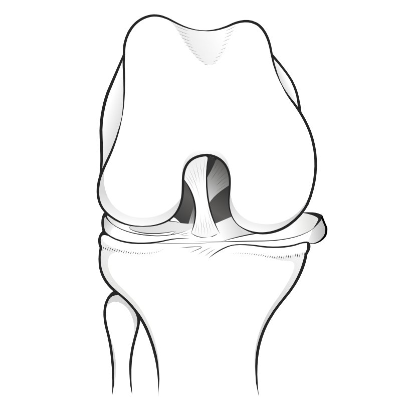
This is the editor's interpretation of a paper published in the orthopaedic literature in 2015 - our attempt to make relevant medical articles accessible to lay readers.
The paper is a review of existing published knowledge up to 2015 regarding meniscal root tears. The authors have reviewed the anatomy, biomechanics and imaging of the meniscal roots, as well as the most common surgical techniques for meniscal root tears and the clinical outcomes. Meniscal root tears can occur chronically in degenerative knees or following acute traumas. Classically, the posterior horns of the menisci bear more load than the anterior horns, especially when the knee is flexed at 90°,15 and are more prone to lesions.
How the menisci protect the knee joint
The authors explain how the menisci (plural of meniscus) function to protect the knee joint from damage and degeneration, by converting the vertical (axial) load going through the knee from the body's weight into a circumferential stress (called 'hoop stress') which is less harmful to the joint surfaces.
At the ends of each meniscus is a root-like extension called the meniscal 'root' which tethers the end of the menisci at the front and the back into the underlying tibial bone. These meniscal roots, they explain, are "fundamental to preserve correct knee kinematics", but it is only in the last 20 years that the importance of the meniscal roots has been understood.
Meniscal roots can tear during an injury but in the medial meniscus the root can tear without any specific injury if the knee is suffering general long-term deterioration. It is usually the roots at the back of the knee - the posterior horns - that are more prone to damage, particularly when the knee is bent to 90°. The medial posterior horn root (at the back of the knee on the inner aspect) is the one that is especially vulnerable. On the lateral side there is more often an association with a knee sprain.
The authors further explain how the meniscus root tear can lead to incompetency of the meniscus "associated with meniscal extrusion, reduced shock absorption, joint degeneration and ultimately osteoarthritis". Meniscal extrusion is when the meniscus slips over the edge of the flattened top of the tibia, and the shock absorbancy is reduced, allowing the bones to come closer together on that side.
Diagnosis
Diagnosing the cause of meniscal incompetence is a challenge in both those cases associated with an injury and those who suffer a tear without a precipitating injury.
In this latter chronic setting, patients may "complain about posterior joint line pain mostly at maximum degrees of flexion or, more rarely, about locking or giving way of the knee."
The authors describe a test by Seil and his colleagues (see references in original document) described a clinical test for medial meniscal root avulsions, where the surgeon feels for the bulge of an avulsion in the joint line when the knee is fully extended and in varus (ie angled outwards).
MRI diagnosis relies on two signs:
- the ghost sign - where - in certail views (sagittal views) - there is nothing in the space where the meniscus should be
- meniscal extrusion - where the meniscus can be seen hanging over the edge of the tibia
The extrusion, they explain, is usually "the result of considerable disruption of the circumferential fibres of the meniscus with loss of the ability to resist hoop strain."
Meniscal extrusion may begin before the actual tear, and measurements may predict when the root is under critical strain. Also the presence of bone marrow swelling in that region may be a harbinger of potential root tear.