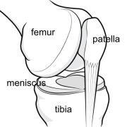
The main bones of the knee joint.
Anatomy means the 'shape' of the structures in the body, and how the different parts correspond to one another. Page updated June 2024 by Dr Sheila Strover (Clinical Editor)
The main bones of the knee joint.
One can see how the bones fit together, but the illustration may leave off the ligaments, tendons and muscles that make the bones functional. So you need to look separately at different illustrations of all the structures to fully understand the functional anatomy.
Doctors have the benefit of having dissected cadavers during their studies - which are, of course three dimensional.
There are two 'tibiofemoral' joints (contact surfaces of tibia and femur), and one 'patellofemoral' joint (contact surface of patella and femur).
The thinner fibula bone at the side of the tibia does not form part of the knee joint itself.
Amongst most important ligaments are the cruciate ligament (inside the capsule) and the collateral ligaments (outside the capsule).