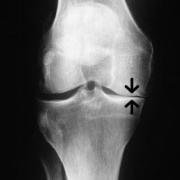
On the one side the normal X-ray 'gap' has gone and bone is touching bone. The normal 'gap' is filled with joint cartilage and meniscus, but on this one side that has worn away.
Bone-on-bone is a term used when joint and meniscus cartilage of the tibiofemoral joint are worn down so much that - instead of nice shiny opposing joint surfaces - exposed bone is grating on exposed bone. Page updated January 2024 by Dr Sheila Strover (Clinical Editor)
On the one side the normal X-ray 'gap' has gone and bone is touching bone. The normal 'gap' is filled with joint cartilage and meniscus, but on this one side that has worn away.
An X-ray finding such as 'bone-on'bone' does not always correlate well with the amount of pain the patient experiences.
"Non-weight-bearing and weight-bearing radiographs of the knee in extension were found to be of limited value in assessing [osteoarthritis] disease status, whereas all standing flexed knee positions reliably imaged joint space width and bone changes in the tibiofemoral joint. Skyline rather than lateral views of the patellofemoral joint were better at detecting joint changes in osteoarthritis