-
The patella - important anatomical relationships
- Pain syndromes related to the patella
- The unstable patella
- Cartilage damage of the patella
- Fractures and tendon ruptures involving the patellar mechanism
What is the patella?
The patella is the kneecap - the bone at the front of the knee.
Strangely the patella is a bone within a tendon - the tendon of the quadriceps muscle ('quads') that spans the region from the hip to the shinbone.
It is very unusual in the body to find a bone inside a tendon (a 'sesamoid' bone), and what it means in reality is that any alteration of the direction of forces in the whole of the quads muscle/tendon/bone complex has to affect the patella.
Therefore many conditions, from the hip down to the foot, may result in pain experienced around the patella.
This is key to understanding problems involving the patella.
The extensor mechanism
The name given to that chain of structures that involves the patella and extends the knee is the 'extensor mechanism'.
Besides the patella, the extensor mechanism includes:
the quadriceps muscles
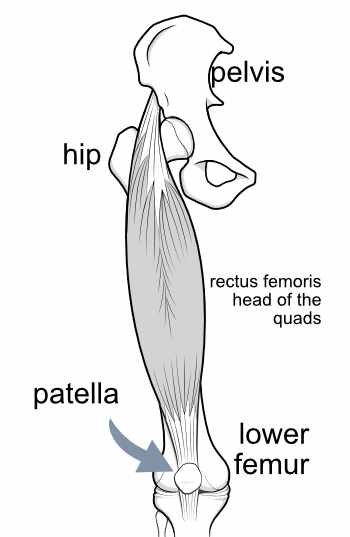
The quadriceps muscle has four muscle heads terminating at its lower end in a single tendon. The largest muscle head is the rectus femoris.
The rectus femoris muscle attaches at its upper end to the pelvis and the top of the femur (thighbone), and at the lower end to the tibia (shinbone) at an important lumpy spot about 1.5 inches below the bottom of the patella - the 'tibial tubercle'. Many procedures to improve patellar pain and stability involve re-locating the tibial tubercle, which basically shifts the alignment of the whole muscle complex.
the quadriceps tendon and the patellar tendon
Because the patella separates the tendon of the quadriceps muscles into two parts, the two parts have separate names. Above the patella the tendon is called the quadriceps tendon and below the patella it is called the patellar tendon (sometimes more correctly called the patellar ligament).
the tibial tubercle
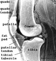
This image on the right (an MRI scan with reverse contrast) shows the knee from the side, with the patella, quadriceps tendon and patellar tendon. The tibial tubercle (or tibial tuberosity) - the bony attachment site of the patellar tendon to the tibia - should align well with the groove where the patella glides and this alignment is calculated from CT or MRI scans as the tt-tg distance which is normally 9mm or less.
the trochlear groove of the femur
The trochlear groove is the groove in the femur bone along which the patella glides when the knee is bent and straightened. You an understand this best if we illustrate it from above, as if you were seated and looking down at your toes. Usually the outer (lateral) wall of the groove is higher than the inner (medial) wall, and the patella is contained within these walls. The undersurface of the patella is v-shaped and snugs into the groove. Although it is free to slide up and down the groove as the leg bends and straightens, it is not free to move from side to side as it is contained by the walls of this groove.
If the groove is insufficiently deep or the walls too low, then the patella may on occasion flip (patellar subluxation) or pop completely out of the groove (patellar dislocation).
Fibrous restraints of the patella
The medial and lateral retinacula
The patella is restrained from moving sideways bytfibrous tethers on each side - the medial and lateral retinaculum. The most significant of these is the lateral retinaculum which may become excessively tight and cause problems.
The medial patellofemoral ligament (MPFL)
On the inner side of the patella another important guy rope is the medial patellofemoral ligament (MPFL) which may become damaged after a patellar dislocation event.
The fat pad
The fat pad is not part of the patellar mechanism but is intimately related to it anatomically. It acts as a fatty cushion protecting the underlying soft tissue structures from impacts to the patella.
Finally, you may like to take a look at this video below, which has been taken during arthroscopic surgery, and which shows the patellafrom above as in the sketch just above the video.
You will see that as the knee is bent by the surgeon the patella engages in the groove of the femur and slips out of view.
[You may have also seen a curtain-like suprapatellar plica stretching horizontally across the field of view on the left. This is a different topic, but mentioned just so that you know. The shadowy stuff on the right is just some unimportant loose tissue floating into view.]
INTRODUCTION: The Patella - a Primer
NEXT PART: Pain syndromes related to the patella