An overview of the anatomy and key issues of damage to the knee meniscus.
 First published in 2011, and reviewed August 2023 by Dr Sheila Strover (Clinical Editor)
The meniscus can be thought of as the shock absorber of the knee, but it also plays an important role in maintaining knee stability.
Pronunciation
Where are the menisci situated?
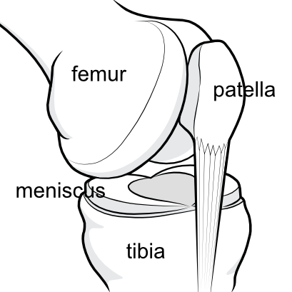
The meniscus on each side of the joint is sandwiched horizontally between the femur (thighbone) and the tibia (shinbone), cushioning the incongruency between the rounded ends of the femur and the flattened top of the tibia.
The meniscus on the inner aspect of the knee is called the medial meniscus and that on the outer aspect is called the lateral meniscus. They have attachments to one another, to the bones and to the capsule, although that is not shown in this simplified illustration.
What does the knee meniscus do?
The knee meniscus helps to stabilise the knee joint and acts as a shock absorber within the knee.
Each meniscus is crescentic in shape with a wedge-shaped cross-section and acts as a spacer between the two long bones of the joint. Not only does the meniscus act as a spacer, but each is filled with fibres which help deflect the vertical forces going through the knee out to the thicker outer rim of the meniscus, so that the more vulnerable joint cartilage of the two long bones is protected from impact. The meniscus also helps to stabilise the joint because of ligament attachments between the menisci, to the bones above and below, and to the capsule surrounding the joint.
Differences between the medial and the lateral meniscus
It is the medial meniscus, on the inner aspect of the knee, that is more commonly injured.
The lateral meniscus is more O-shaped and quite highly mobile, able to slide forwards and backwards with knee movement. In addition there is a tendon called the popliteus tendon that passes along one edge, which further breaks the attachment of the meniscus rim to the capsule of the joint at that point, and this adds to the mobility of the lateral meniscus.
The medial meniscus is larger and more C-shaped, and is more tightly bound to the capsular structures and to the medial collateral ligament along its outer rim. It moves very little with rotational movement of the knee. It is this tethering which leads to the medial meniscus being torn more frequently than the lateral meniscus. The lateral one can move and absorb impact, while the medial one simply rips.
 |
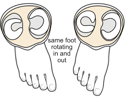 |
| Because of its particular anatomy it is the medial one that is more commonly injured. | Here you can see that when the lower limb is rotated, the smaller, rounder lateral meniscus moves more than the medial. |
Anchors of the meniscus
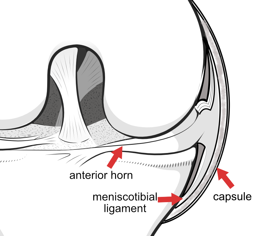
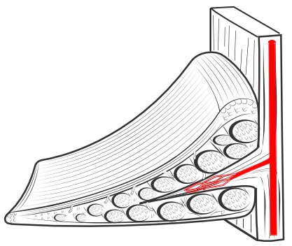
The ends of the meniscus at the back and front of the knee are called the horns. The horns are tethered to the underlying bone via a meniscal root, which extends down at right angles under the horn.
The horn at the back is the posterior horn and the one at the front is the anterior horn. The middle bit of the meniscus is called the body of the meniscus. The meniscus body is wedge-shaped, with the thicker lateral rim on the outer side and the flattened medial rim on the inner side. Both menisci anchor to the tibia via the meniscotibial ligament. The back of the lateral meniscus also attaches to the femur via the meniscofemoral ligament, which splits into two parts, one going in front of the posterior cruciate ligament one going behind it. Behind the lateral meniscus, a meniscofibular ligament connects the meniscus to the fibula bone. The outer rim of both menisci have loose connections to the capsule, which are more complete on the medial side.
The important meniscus fibres
A cross-section of the wedge-shaped meniscus shows that it is packed with bundles of fibres, both longitudinal and horizontal. These fibres are embedded in a cartilagenous matrix, and it is the combination of fibres and matrix that give the meniscus its strength and flexibility.
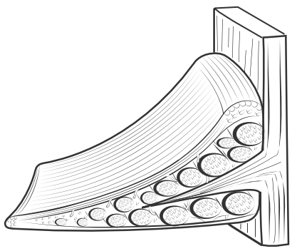
The fibres are arranged so as to deflect the vertical forces passing through the meniscus, so that the stress is taken on the outer rim of the meniscus - this is called 'hoop stress', and if the fibres become disrupted then the underlying joint cartilage of the tibial comes under much greater stress than normal.
Meniscus blood supply
The blood supply of the meniscus enters from the outer rim. Very little blood reaches the inner rim which is nourished only by the joint fluid inside the knee. This is important in healing of meniscus tears and tears in the inner rim do not heal well.
Surgeons refer to tears in the outer rim as being in the red-red zone, which is a reference to the good blood supply there. Tears of the inner rim are referred to as being in the white-white zone, and tears between these areas are referred to as being in the red-white zone.
A note about terminology
When a sportsperson says "I have torn my cartilage", what he should be saying is "I have torn my meniscus". You see, anatomists used to call the knee menisci 'semi-lunar cartilages' (which means 'half-moon-shaped' cartilages), and the 'semi-lunar' bit was often dropped.
However, the gristle at the ends of the long bones is also covered in a material which is called 'hyaline cartilage', and when it became clear that patients were getting muddled about the two different structures both tagged 'cartilage', doctors began calling the one 'meniscus' (previously the semi-lunar cartilage) and the other 'cartilage' (meaning hyaline cartilage).
Unfortunately, the confusion over the terms still persists.
So, to avoid confusion -
- Drop the plural term 'cartilages' completely
- Refer to 'menisci' if you mean the shock absorbers between the femur (thighbone) and tibia (shinbone)
- Refer to 'cartilage' when you mean the hyaline cartilage at the end of the long bones (the 'gristle')
- Don't let the surgeon patronise you by using the old terminology and referring to your 'cartilages' when he really means your 'menisci'. Ask him to clarify the terminology or to draw a picture.
- Try not to be confused by the many internet articles still muddling up the old and new terminology.
INTRODUCTION TO COURSE: Key issues relating to the knee meniscus
NEXT PART: Meniscus tears
