Dr Noyes explains the various considerations of the rehab team as you go through rehabilitation after posterolateral corner surgery.
 First published 2009 by Dr Frank R Noyes, and reviewed August 2023 by Dr Sheila Strover (Clinical Editor)
Rehabilitation protocol after posterolateral corner surgery - course
- Rehabilitation after posterolateral corner surgery
- Rehabilitation protocol after posterolateral corner surgery
- Rehabilitation protocol after posterolateral corner surgery - Phase 1. Weeks 1-2 (Visits: 2-4)
- Rehabilitation protocol after posterolateral corner surgery - Phase 2. Weeks 3-4 (Visits: 2-4)
- Rehabilitation protocol after posterolateral corner surgery - Phase 3. Weeks 5-6 (Visits: 1-2)
- Rehabilitation protocol after posterolateral corner surgery - Phase 4. Weeks 7-8 (Visits: 1-2)
- Rehabilitation protocol after posterolateral corner surgery - Phase 5. Weeks 9-12 (Visits: 1-2)
- Rehabilitation protocol after posterolateral corner surgery - Phase 6. Weeks 13-26 (Visits: 2-3)
- Rehabilitation protocol after posterolateral corner surgery - Phase 7. Weeks 27-52 (Visits: 2-3)
Related course: Posterolateral corner injuries of the knee
The rehabilitation program described in this section consists of a careful incorporation of exercise concepts supported by scientific data and clinical experience (see the reference list below).
The goal is to progress a patient through the exercise program on a rate that takes into account sports and occupational goals, condition of the knee joint (articular cartilage surfaces and arthritic damage) and menisci, return of muscle function and lower limb control, postoperative healing, and graft remodeling.
Modifications to the postoperative exercise program may be required if noteworthy arthritis is found during surgery. In these patients, a return to very strenuous high-impact activities (such as jumping, twisting, turning, cutting, pivoting) is not recommended. The program is used after all of the posterolateral procedures I described in Part 5 of this course, including anatomic and proximal advancement techniques of the posterolateral structures, and nonanatomic femoral-fibular reconstruction.
The supervised rehabilitation program is supplemented with home exercises performed daily. In my Center, our therapists routinely examine the patients in the clinic in order to progress the protocol in a safe and effective manner. Therapeutic procedures and modalities (such as electrical muscle stimulation and biofeedback) are used as required.
Avoiding complications
Our therapists and surgeons continually monitor the patients for joint swelling, pain, gait pattern, knee motion, patellar mobility, muscle strength, and flexibility after surgery. We warn patients at the 4th to 8th postoperative weeks that, as they resume weight bearing and wean off of crutches, to avoid a varus or internal tibial rotation position which could place high forces on the posterolateral structures. It is important that the patient demonstrates good lower extremity muscle control and a normal walking pattern before crutches are discontinued.
Patients begin protected knee motion exercises the day following surgery, but we use maximal protection against high joint loads to prevent stretching and failure of posterolateral grafts or advancement procedures. Patients are warned to avoid hyperextension and activities which would incur varus loading on the joint. Delays in return of full knee flexion, full weight bearing, initiation of certain strengthening and conditioning exercises, and running and return to full sports activities are incorporated.
Posterolateral reconstructions are monitored after surgery with manual examination of lateral joint opening and external tibial rotation. Lateral stress x-rays may be used if an early increase in these knee motions are detected. Any patient who has problems progressing through the protocol or who develops a complication requires additional supervision in the formal clinic setting.
Rehabilitation program
After surgery, the patient’s leg is placed into a bi-valved cast that is used for the first 4 postoperative weeks to provide maximum protection to the knee joint and posterolateral structures. “Bi-valved” means that the cast is cut so that it can be removed 4 times a day for passive knee motion exercises which are performed in a seated position with a 10-pound valgus load applied to decrease lateral joint forces. At 4 weeks postoperative, the cast is removed and replaced with a hinged brace that is locked at 10° of flexion. The brace is removed 4 times each day for range of motion exercises. At 6 weeks postoperative, the brace is unlocked as knee flexion to 110° is encouraged and partial weight bearing allowed.
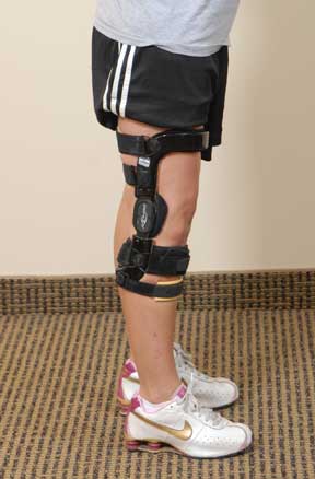
At 7-8 weeks, a custom medial unloading brace (see Figure on right) is applied as weight bearing progresses to full and flexion is advanced to 120°. The brace is also used as patients return to activity to provide protection against knee hyperextension and excessive varus loads.
Patients are allowed 0° to 90° of knee motion immediately postoperatively. Flexion is slowly advanced to 110° by postoperative week 5, 120° by week 8, and 130° by week 12. Patients are cautioned to avoid varus tensioning when performing knee flexion exercises. They are taught (and the assistance of a partner is encouraged) to place a hand on the lateral (outside) portion of the knee and create a 10-pound valgus load to protect the posterolateral structures.
Patients are not allowed to bear weight on their operated leg for the first 2 postoperative weeks. Then, partial weight bearing (25% of the patient’s body weight) is begun at postoperative weeks 3-4, with slow advancement to full by week 8 with cane or crutch support. A cane or crutch is used for approximately another 3 to 4 weeks if needed. I want to see the patient walk normally and have good muscle control before we discontinue the walking support aids.
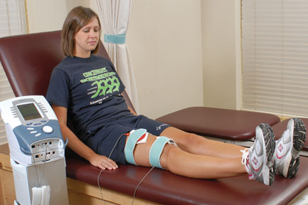
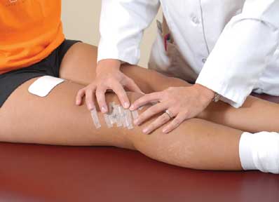
Patellar mobilization (see Figure below), flexibility exercises, electrical muscle stimulation (see the Figure on left), cryotherapy, biofeedback, and the strengthening and conditioning programs are done as shown in the Tables in Part 7.
[This article was published in Noyes’ Knee Disorders: Surgery, Rehabilitation, Clinical Outcomes, Noyes FR, Barber-Westin SD (eds.), Copyright Saunders, 2009 - Noyes FR, Barber-Westin SD, Heckmann TP: Rehabilitation of posterior cruciate ligament and posterolateral reconstructive procedures, pages 631-657.]
In certain athletes, a running program is begun at approximately the 9th postoperative month, and plyometric and sports specific training programs are initiated at the 12th postoperative month. However, the majority of patients who require major posterolateral ligament reconstructive procedures do not desire to return to high-impact sports and therefore, this advanced conditioning and training is usually not required. Patients who have arthritic damage are advised to return to low-impact activities only to protect the knee joint.
Discharge criteria following lateral and posterolateral graft reconstructions is based on patient goals for athletics and occupation and the rating of symptoms, lateral joint opening, muscle strength testing, and function testing.
References
1. Grood, E. S.; Stowers, S. F.; and Noyes, F. R.: Limits of movement in the human knee. Effect of sectioning the posterior cruciate ligament and posterolateral structures. J Bone Joint Surg Am, 70(1): 88-97, 1988.
2. Noyes, F. R., and Barber-Westin, S. D.: Posterolateral knee reconstruction with an anatomical bone-patellar tendon-bone reconstruction of the fibular collateral ligament. Am J Sports Med, 35(2): 259-73, 2007.
3. Noyes, F. R., and Barber-Westin, S. D.: Surgical reconstruction of severe chronic posterolateral complex injuries of the knee using allograft tissues. Am J Sports Med, 23(1): 2-12, 1995.
4. Noyes, F. R., and Barber-Westin, S. D.: Surgical restoration to treat chronic deficiency of the posterolateral complex and cruciate ligaments of the knee joint. Am J Sports Med, 24(4): 415-26, 1996.
5. Noyes, F. R.; Cummings, J. F.; Grood, E. S.; Walz-Hasselfeld, K. A.; and Wroble, R. R.: The diagnosis of knee motion limits, subluxations, and ligament injury. Am J Sports Med, 19(2): 163-71, 1991.
6. Noyes, F. R.; Dunworth, L. A.; Andriacchi, T. P.; Andrews, M.; and Hewett, T. E.: Knee hyperextension gait abnormalities in unstable knees. Recognition and preoperative gait retraining. Am J Sports Med, 24(1): 35-45, 1996.
7. Noyes, F. R., and Grood, E. S.: Diagnosis of knee ligament injuries: Five concepts. In The Crucial Ligaments. Edited by Feagin, J., New York, Churchill Livingstone, 1988.
8. Noyes, F. R.; Schipplein, O. D.; Andriacchi, T. P.; Saddemi, S. R.; and Weise, M.: The anterior cruciate ligament-deficient knee with varus alignment. An analysis of gait adaptations and dynamic joint loadings. Am J Sports Med, 20(6): 707-16, 1992.
9. Noyes, F. R.; Stowers, S. F.; Grood, E. S.; Cummings, J.; and VanGinkel, L. A.: Posterior subluxations of the medial and lateral tibiofemoral compartments. An in vitro ligament sectioning study in cadaveric knees. Am J Sports Med, 21(3): 407-14, 1993.
INTRODUCTION TO COURSE: Rehabilitation protocol after posterolateral corner surgery - course
NEXT PART: Rehabilitation protocol after posterolateral corner surgery
Dr Frank R Noyes

DR NOYES HAS RETIRED.
Based at the Cincinnati Sportsmedicine and Orthopaedic Center in the USA, Dr Frank Noyes is one of the world's most prominent figures when it comes to knee surgery. A prolific researcher and writer, he has published over 200 studies and articles in the world's top...read more