Published 2007

A Poem

The history, having told you what you needed

To know of symptoms, you have then proceeded

To look and feel and move the painful knee

To postulate the main pathology.

But now your diagnostic postulations

Require the help of more investigations.

Foremost in these modern special tests

Are some essential imaging requests.

ap x-ray of knee

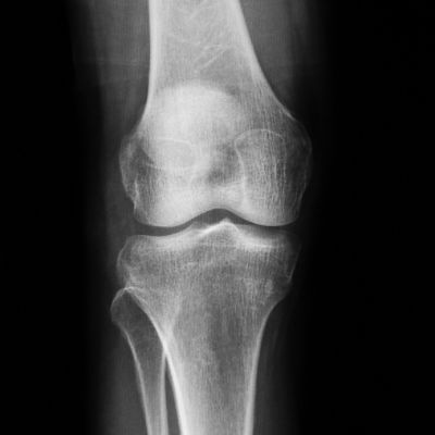
With radiographic pictures and C.T.

The bones are clearly shown, and you can see

The length, alignment and, to some extent,

You can judge the mineral content.

Alignment of the bones whilst bearing weight

Is better judged while the knees are not quite straight.

Fifteen degrees, or more, of flexion now

Accentuates the wear and tear somehow.

When bone-on-bone is what you want to capture

the "Rosenberg’s view” will send you into rapture.

knee bones from the side

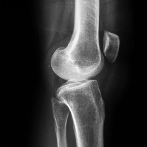
In the lateral it is most important too

That both the condyles must be shown with true

Superimposition - one upon the other -

(Otherwise you need not even bother.)

The lateral and PA views erect,

Whilst good, are yet not able to project

The better details of patellar tracking

As for these you need the further backing 

Of skyline views in some degrees of flexion

To document the tracking to perfection.

To measure trochlear depth you must be sure

To obey the rules of Henri Dejour.

The trochlea seen in truly lateral view

Shows one profile, but an extra shadow too.

This shadow is the bottom of the "V"

Which quite clearly in the skyline views you see.

The depth of this most important groove

Determines how the kneecap's going to move,

And whether or not it has the capability

To develop patellofemoral instability.

Whilst thinking now of X-rays or C.T

The latter has advantages you’ll see

In considering the planning for correction

Of length and alignment to perfection.


-