Articular cartilage has a very unique structure with few cells and a high water content.
 Page updated July 2023 by Dr Sheila Strover (Clinical Editor)
This Primer explains how joint (articular) cartilage damage can lead to osteoarthritis in the knee.
- Introduction to arthritis
- Types of arthritis
- The common arthritis pathway
- How knee arthritis progresses
-
What's special about hyaline (articular) cartilage?
- Arthritis cysts and spurs
- Joint injections for knee arthritis
- The concept of arthritis compartments
- Classifying the amount of cartilage damage
- Knee X-rays and arthritis
If one looks at the microscopic structure of cartilage one will see that it is composed primarily of a slippery white 'matrix' containing few cells and strengthened by the incorporation of randomly arranged elastin fibres.

The matrix has a high water content, a fairly high collagen content (a structural protein) and a percentage of something called proteoglycans (including chondroitin and keratin sulphate) which are responsible for attracting the water into the cartilage and maintaining its compressive strength.
Within the matrix are a relatively few cartilage cells ('chondrocytes') suspended, often quite apart from one another, within small 'bubbles' called lacunae.
Where the cartilage layer joins onto the bone beneath it is a region known as the 'tidemark'.
Tidemark
If the cartilage is lacerated, cuting through the tidemark and into the sub-chondral bone then some healing can occur, although the cartilage that forms is 'fibrocartilage' rather than true joint (or 'articular') cartilage. This can occur because stem cells from the bone marrow can migrate into the blood clot that forms and these cells can mature into fibrocartilage. This type of cartilage is weaker than the original cartilage, but better than nothing.
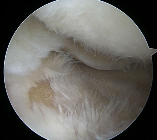
If there is a split but it does not go through the tidemark, there is no healing, and gradually the surface will become friable, and fibrillated, and the cartilage will wear away down to the bone, as you can see in this photograph which has been taken during keyhole surgery (courtesy Prof Adrian Wilson).
Osteoarthritis
Cartilage breakdown is the first stage of osteoarthritis. Initially the process may be painless, but pain will become a feature once the underlying bone becomes exposed.
PREVIOUS PART: How knee arthritis progresses
NEXT PART: Arthritis cysts and spurs
