The trochlear groove is the concave surface where the patella (kneecap) makes contact with the femur (thighbone). Also called the 'trochlea'.
 Page updated March 2024 by Dr Sheila Strover (Clinical Editor)
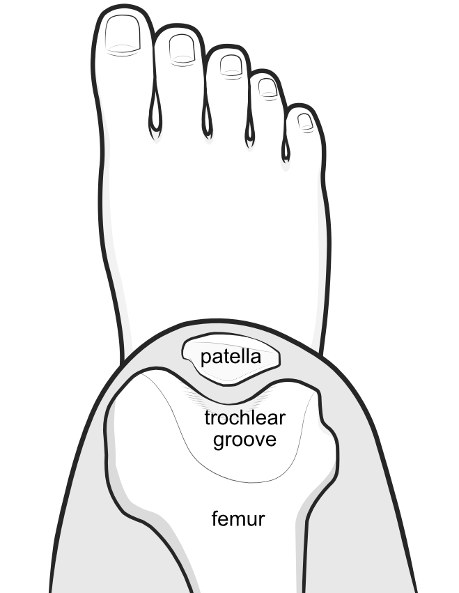
As the knee bends, the patella engages with the groove and is contained by its steep walls.
The pulley mechanism within the trochlear groove
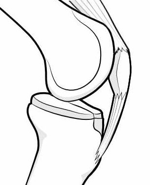
The lower part of the patella is tethered to the tibia bone, while the upper part is continuous with the big quadriceps muscle group that makes up the 'lap'. When the quadriceps (quads) contract and relax, as it does when walking, the joint straightens and bends, but the patella normally remains restrained within the groove during that excursion, acting as a 'pulley' to increase the force..
If the groove for some reason is not 'doing its job' then the patella may ride up, and even over, the walls of the groove - this is called 'subluxation' and 'patellar dislocation', and it may happen when the upper part of the groove is anatomically abnormal (trochlear dysplasia), if the patella itself is anatomically abnormal (patellar dysplasia) or if the patella is situated too high in the groove (patella alta).
Evaluating the trochlear groove in patellar instability
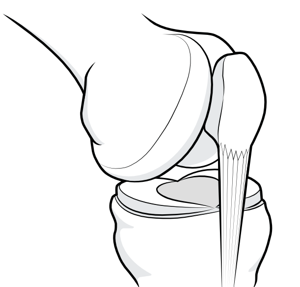
Towards the top of the trochlear groove the walls are flatter. When the knee is in extension (straight) and the patella in the upper part of the groove, it is more easily able to disengage (sublux or dislocate).
Patellar instability may occur with activities or the examining practitioner may be able to'pop the kneecap out' when the knee goes into extension during an examination. There may also be a 'J-sign' where the abnormal excursion of the patella is visible when the patient holds the limb over the edge of the examination couch and flexes and contacts the knee.
Several imaging methods may allow objective measurements of trochlear dysplasia and patella alta - for example, the merchant view X-ray, the lateral trochlear inclination angle and the calculation from CT, MRI or X-ray of the TT-TG distance.
-
Quote from peer-reviewed paper:
"Imaging is essential in the assessment of patellar instability..."
Citation:Batailler C, Neyret P. Trochlear dysplasia: imaging and treatment options. EFORT Open Rev. 2018 May 21;3(5):240-247. doi: 10.1302/2058-5241.3.170058. PMID: 29951262; PMCID: PMC5994618.
Forum discussions
- Please help a newbie!
A patient with a shallow trochlea tries to understand the issues of potential surgery.
Relevant material -
eBook - Factors contributing to an Unstable Patella
by Dr Lars Blønd (Knee Surgeon). Free to download.