Suprapatellar is a positional adjective describing something to be 'above the patella' (kneecap).
 Page updated November 2023 by Dr Sheila Strover (Clinical Editor)
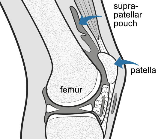
This illustration shows the knee cut through the middle. The large cavity (dark grey) extends above (suprapatellar pouch), behind and below the patella. A small suprapatellar plica is present but it does not form a complete septum so the whole cavity is open to the joint..
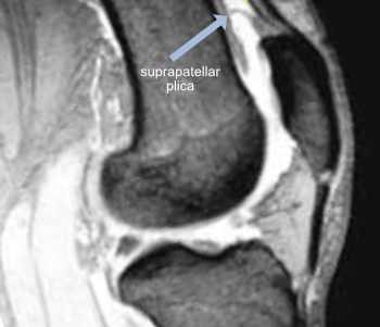
This MRI shows a similar view where the arrow is pointing to a complete suprapatellar septum dividing the cavity into two - the closed bursa (above) and a small pouch below. This may confuse the novice surgeon during arthroscopy.
The suprapatellar pouch during arthroscopy
During keyhole surgery, a cannula is inserted into the knee joint and fluid is pumped in under pressure to inflate the cavity so that the surgeon can see and access the structures of the joint. Both the camera and various instruments may be inserted into the expanded space, allowing surgery to the ligaments, joint surfaces and menisci.
Quick links
Peer-reviewed papers
-
Quote:
"By the fifth month of fetal life there is a suprapatellar septum between the knee joint cavity and suprapatellar bursa that later perforates and involutes so that it established a normal communication between the knee and bursa cavity...A small part of embrionic remnants can later persist as a suprapatellar plica"
Citation: Crnković T, Gašpar D, Ethurović D, Podsednik D, Slišurić F. New insights about suprapatellar cyst. Orthop Rev (Pavia). 2012 Jan 2;4(1):e9. doi: 10.4081/or.2012.e9. Epub 2012 Feb 16. PMID: 22577510; PMCID: PMC3348697.
 2008 -