The lateral retinaculum is the name given to the sheet of fibrous tissue that provide a passive restraint to the patella on the outer (lateral) side of the knee.
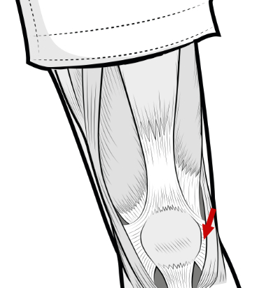
The colours show the superficial and deep layers of the retinaculum. The red arrow marks the lateral retinaculum. On the other side of the patella is the medial retinaculum.
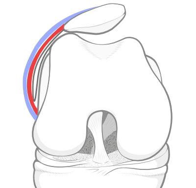
The opposite knee showing patellar tilt due to a tight lateral retinaculum on the lateral side. We have not drawn the retinaculum on the medial side.
Patellar tilt due to a tight lateral retinaculum
It may happen that the lateral retinaculum becomes tight, tilting the patella over to one side. In this situation, the procedure of lateral release or lateral retinacular lengthening may be beneficial.
-
See also -
Peer-reviewed paper -
- 2016 - Anatomy of the lateral retinaculum of the knee. Authors: Merican AM and Amis AA. - and summarised by Dr Sheila Strover (Clinical Editor)